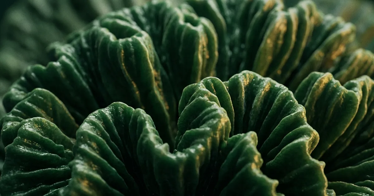
Most people have seen thousands of images of the brain and never seen one. The models in classrooms are rubbery and beige. The renders online are glossy and grey. Neither is quite right, and the differences are more interesting than you would expect.
It is much softer than you think
Start here, because it surprises nearly everyone.
A fresh, unfixed human brain has roughly the consistency of firm jelly, or a very soft cheese. Lifted out of the skull it slumps and deforms under its own weight. It cannot hold its own shape.
The specimens people handle in teaching labs have spent weeks in formalin, which cross-links the proteins and turns the tissue rubbery and self-supporting. That is why anatomy specimens can be picked up and turned over. It is also why the thing most of us picture as a real brain is in fact a chemically altered version of one.
The colour
A living brain is pinkish grey and glossy, not the flat beige of a preserved specimen. It is wrapped in membranes, the meninges, and threaded with fine red blood vessels running across the surface.
The grey matter of the cortex is only the outer two to four millimetres. Slice into it and the interior is paler and creamier. That is the white matter, and its colour comes from the fatty myelin insulating the connecting fibres. Grey matter and white matter are named for exactly what they look like on a cut surface.
Size and weight
An adult human brain weighs roughly 1,300 to 1,400 grams, about three pounds. Classic measurements put the average at around 1,336 grams for adult men and 1,198 grams for adult women, a difference that tracks overall body size and predicts nothing about ability.
For a physical comparison: about the weight of a large cabbage, or a small laptop. It fits in two cupped hands.
Inside the skull it does not weigh that, because it floats. Suspended in cerebrospinal fluid, its effective weight drops to something closer to 50 grams. This is not a curiosity. Without that buoyancy the underside would be crushed by the tissue above it.
The folds
The wrinkles are the most recognisable feature and the most misunderstood. They are not decoration and they are not a sign of intelligence in individuals.
The cortex is a sheet. Unfolded, it covers roughly 2,500 square centimetres, about the area of a large table napkin or an open newspaper page. Fitting that into a skull the size of a grapefruit requires folding it, and the ridges and valleys are the result. Species with smaller cortical sheets relative to skull size have smoother brains.
What you cannot see
Look at a real brain and the regions you have read about are simply not visible as separate parts. There is no line where the frontal lobe ends and the parietal begins.
The lobes are named after the skull bones above them. Their boundaries are drawn along convenient landmarks, the deep grooves called sulci, and those grooves vary from person to person. What looks on a diagram like a border between countries is, in the tissue, a fold that anatomists agreed to treat as a boundary.
The one division you can see immediately is the split between the two hemispheres, and the cerebellum at the back, which has a visibly different texture: fine, tightly packed parallel ridges rather than the broad folds of the cortex. Our interactive map of the human brain shows how the named regions map onto that continuous surface.
Where to actually see one
Medical museums are the realistic option. The Mütter Museum in Philadelphia holds a well-known collection, and many university anatomy departments maintain specimens that are open to the public or to visiting groups.
Buying one is not a route. Trade in human remains is restricted or outright illegal in most countries, and material offered for sale is frequently either not human or not legally obtained.
If you are searching for photographs, the useful thing to know is which kind you are looking at. Preserved specimen, fixed and pale. Surgical photograph, pink and glossy and wet. Plastinated display, colour-enhanced and stiffened. They are three quite different objects, and only one of them looks like what is currently inside your head.
Why the gap between model and object is worth noticing
There is a lesson buried in that mismatch, and it is not really about brains.
Nearly everyone who pictures a brain is picturing a teaching model: firm, beige, colour-coded, with the lobes helpfully divided. Every one of those properties was added by someone trying to make the thing easier to learn from. The real object is soft, undivided, and one colour.
That is how almost all learning starts. You are handed a simplified version, it works, and then at some point the simplification quietly becomes the thing you believe. The model brain has borders because a diagram needs borders. The real one does not, and the difference matters the moment anyone tries to sell you something based on which part of your brain you use.
Worth carrying past this page: when a fact feels unusually tidy, it is often the teaching version. That is not a reason to distrust it. It is a reason to ask what got smoothed off.
Frequently Asked Questions
What does a real human brain look like?
Pinkish grey and glossy, wrapped in membranes and threaded with fine red blood vessels. The surface is deeply folded. It is soft enough that a fresh, unpreserved brain deforms under its own weight when lifted out of the skull.
How much does a human brain weigh?
Roughly 1,300 to 1,400 grams in an adult, about three pounds. Averages are around 1,336 grams for adult men and 1,198 grams for adult women, a difference that tracks overall body size.
How soft is a real brain?
About the consistency of firm jelly or very soft cheese. The rubbery, self-supporting specimens used for teaching have been hardened in formalin for weeks, which also drains their colour.
Why does a brain look beige in photographs?
Because most photographs show preserved specimens. Formalin fixation both stiffens the tissue and removes much of the colour. A living brain is pinker and glossier than the preserved version most people picture.
Where can you see a real human brain?
Several medical museums display genuine specimens, including the Mütter Museum in Philadelphia and various university anatomy collections. Buying human remains is restricted or illegal in most countries, so museums are the realistic route.
Every NerdSip course clears a four-layer fact-checking pipeline before it reaches the app: grounded in live sources, scored nightly, gated on failure, and reviewable by a person at one tap. Articles on this blog are drafted with AI assistance, then researched, verified, and edited by our team.
Editorial responsibility: ai51 UG (haftungsbeschränkt). Responsible editor named in the imprint.
📚 Keep Learning
- How Many Brain Cells Does a Human Have?
- What Is the Primary Function of the Human Brain?
- The Main Regions of the Human Brain, Explained
- When Does the Human Brain Fully Develop?
- 27 Mind-Blowing Brain Facts That Will Change How You Think About Thinking
- Neuroplasticity: How Your Brain Rewires Itself (At Any Age)
Explore the NerdSip learning hubs
The blog is our editorial layer. When you want to move from reading into doing, these hubs organize the rest of NerdSip by format, including the app itself and the press that has written about it.
Learn one real thing today
NerdSip turns any topic you type into a fact-checked five-minute course, brain science included. Free to start.
4.7 stars from 380 reviews · 8,900+ learners